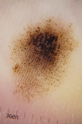
Teledermatology or the practice of Dermatology with the use of digital images, has come a long way since its inception in the early 1990’s. Initial studies were limited by poor quality images and slow internet speeds. The role of teledermatology is ever increasing now with advancements in technology.
Recent studies have shown that when provided with dermoscopic images (Dermoscopy is the use of a Dermatoscope, a polarising microscope), Dermatologists were able to diagnose skin lesions with the same accuracy as a face-to-face consultation.1-4 In fact, the author has published one of the pioneering articles on this topic.4
However, there are several points to consider when deciding between a face-to-face examination versus a “scan”:
- Who is viewing the images? – Published studies have compared the accuracy of teledermatology when the images are read by Dermatologists. We have insufficient evidence for images that are interpreted by other specialties. MoleMap® is a system of digital dermoscopy where the images are viewed by Dermatologists. Before engaging in a “scan”, it is important to enquire who is visualising the image? Is it a Dermatologist or is it some other practitioner?
- Who is taking the image? – Is it a doctor or a technician?
- Which lesion will be imaged? – Most digital photography systems image the lesion that you are concerned about, or the lesion that they spot. However, you could potentially have a suspicious lesion that neither you nor the person imaging the lesion have noted.
In summary, it is always appropriate to have a first face-to-face skin check with a Dermatologist. A skin check is not just a check of your moles. It is a full examination of your skin including non-pigmented lesions and will also involve the use of a dermatosope – a similar tool to what is involved in teledermatology above. It is not uncommon that Dermatologists pick up spots that you are not aware of. A skin check also involves risk stratification, skin care and sun protection advice. Your Dermatologist may then recommend a follow up face-to-face examination or a teledermatology follow up. This decision is dependent on your skin type, the number of lesions you have and the risk of skin cancer.
References
- Blum A, Luedtke H, Ellwanger U, Schwabe R, Rassner G, Garbe C. Digital image analysis for diagnosis of cutaneous melanoma. Development of a highly effective computer algorithm based on analysis of 837 melanocytic lesions. Br J Dermatol.2004 Nov;151(5):1029-38.
- Fabbrocini G, Balato A, Rescigno O, Mariano M, Scalvenzi M, Brunetti B. Telediagnosis and face-to-face diagnosis reliability for melanocytic and non-melanocytic ‘pink’ lesions. J Eur Acad Dermatol Venereol.2008 Feb;22(2):229-34. doi: 10.1111/j.1468-3083.2007.02400.x.
- Piccolo D, Smolle J, Wolf IH, Peris K, Hofmann-Wellenhof R, Dell’Eva G, Burroni M, Chimenti S, Kerl H, Soyer HP. Face-to-facediagnosis vs telediagnosis of pigmented skin tumors: a teledermoscopic study. Arch Dermatol. 1999 Dec;135(12):1467-71.
- Tan E, Yung A, Jameson M, Oakley A, Rademaker M. Successfultriage of patients referred to a skin lesion clinic using teledermoscopy (IMAGE IT trial). Br J Dermatol. 2010 Apr;162(4):803-11. doi: 10.1111/j.1365-2133.2010.09673.x. Epub 2010 Mar 5.


